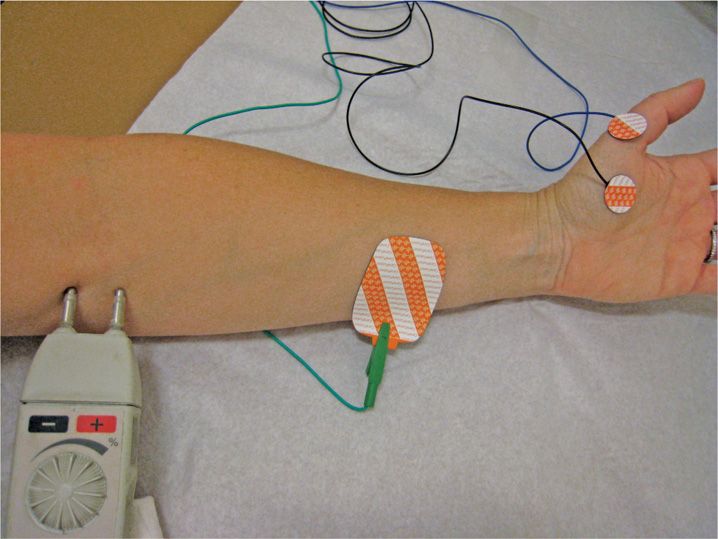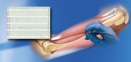
The recording should show no electrical activity when the muscle is at rest. It may also be heard on a loudspeaker as machine gun-like popping sounds when you contract the muscle. The electrical activity in the muscle shows as wavy and spiky lines on a special video monitor. The needle may be moved several times to record the electrical activity in different areas of the muscle or in different muscles. Then the technologist or doctor asks you to tense the muscle with gradually increasing force while the electrical activity in the muscle is recorded. Once the electrodes are in place, the electrical activity in that muscle is recorded while the muscle is at rest. The electrode is attached by wires to a recording machine. You will feel a brief, sharp pain when a needle electrode is inserted into the muscle.

An electrode that combines a reference point and a needle for recording is inserted into the muscle. Typically, the skin over the area being tested will be cleaned with an antiseptic. You will be asked to lie on a table or sit in a reclining chair so that the muscles being tested are relaxed and easy to reach. You may be given a hospital gown to wear.

Wear loose-fitting clothing that permits access to the muscles and nerves to be tested.

Problems in a muscle, the nerves controlling a muscle, the spinal cord or the brain can all cause these kinds of symptoms.Įlectromyograms are useful in determining whether there has been pressure on a nerve or nerve root degeneration.

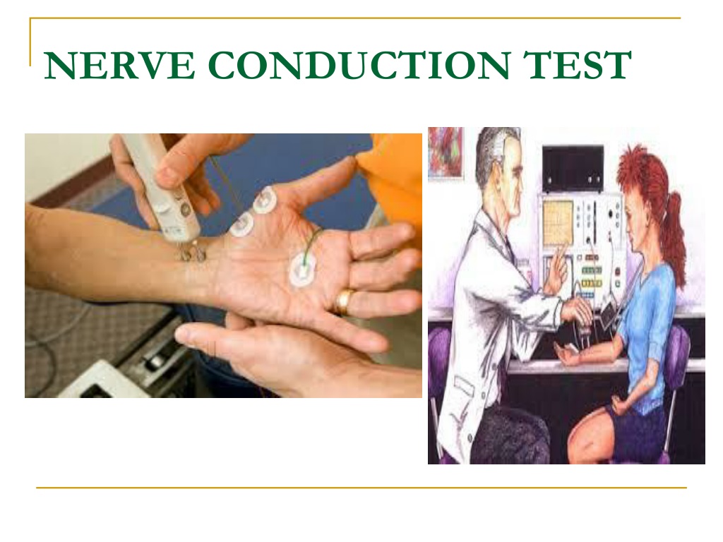
Electromyography is usually done with nerve conduction studies. Electromyography measures the electrical impulses of the muscles at rest and when contracted.


 0 kommentar(er)
